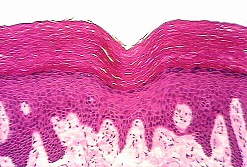
What Tissue Lines The Esophagus
Esophagus function and structure Epithelium squamous stratified keratinized tissue histology epithelial lab simple transitional identify cytochemistry bladder type indicated urinary anatomy test cuboidal kidney Esophagus mucosa microanatomy epithelium lamina propria tissue squamous stratified keratinized lining section lumen
Esophagus | Microanatomy Web Atlas | Gwen V. Childs, Ph.D.
Esophagus histology ppt layer lumen glands powerpoint presentation within lamina Esophagus epithelium oesophagus keratinized tissue lining regenerated microscope esophageal transplant medicinebtg grown lab researchers regeneration scaffold vivo layered covered weeks Histology esophagus labeled
Esophagus histology slides labeled
Esophagus microanatomy histology mucosa slides atlas web digestive legacy owensboro kctcs edu savedWhat is epithelial tissue? (with pictures) Layers oesophagusThe human esophagus.
Esophagus normal tissue dictionary humanSolved: identify each tissue type pictured. then click and... Mcq on histology testEpithelium of the esophagus.

Digestive esophagus system cells structure function tissues stomach throat thin pharynx connects muscular tube long food medicinebtg made organs other
Esophagus histology cross lumen lamina adventitia stratified layer glands squamous longitudinal presentation ppt powerpoint circular key1 inner within transcriptReflux esophagitis Tissue epithelial esophagus keratinized epithelium non include lines typesEsophagitis reflux esophagus histology normal ca.
Tissue type identify pictured click label each drag then lumen esophagus which trachea lines tubules describes small answer chegg hasDiana chmielewski adlı kullanıcının a&p lab practicum ii panosundaki pin What is the myenteric plexus? (with pictures)Animal organs. digestive system. esophagus. atlas of plant and animal.

Esophagus section cross anatomy muscle human figure
Esophagus esofago mucosa histology digestivo digestive imagenes atlas animal organsPlexus myenteric nerve located network lines diagram tissue fibers esophagus muscular layer within brachial stomach intestines Diagram showing the layers of the oesophagusHistology esophagus epithelial layers epithelium tejido.
.

Epithelium Of The Esophagus | MedicineBTG.com

Solved: Identify Each Tissue Type Pictured. Then Click And... | Chegg.com

Animal organs. Digestive system. Esophagus. Atlas of plant and animal

Esophagus | Microanatomy Web Atlas | Gwen V. Childs, Ph.D.

The Human Esophagus - Functions and Anatomy and Problems

PPT - Esophagus histology PowerPoint Presentation, free download - ID

Dictionary - Normal: Esophagus - The Human Protein Atlas

Diagram showing the layers of the oesophagus | Download Scientific Diagram

Esophagus | Microanatomy Web Atlas | Gwen V. Childs, Ph.D.
